What Is the Difference Between Varicose Veins and Spider Veins?
- Advanced Medical & Diagnostic Center
- August 9, 2026
- 10 min read
Vein treatment
Table of Contents
- 1. Why the Distinction Matters
- 2. What Varicose Veins Are
- 3. What Spider Veins Are
- 4. Varicose Veins vs Spider Veins: Key Differences
- 5. What Causes Each Type
- 6. Which One Requires Medical Treatment
- 7. Treatment Options for Each Type
- 8. Frequently Asked Questions
- 9. What to Do If You Are Unsure Which Type You Have
If you have noticed unusual veins on your legs, you have probably wondered whether what you are seeing is serious or simply cosmetic. The terms varicose veins and spider veins are often used interchangeably, but they are different conditions with different causes, different symptoms, and different treatment implications. Understanding the distinction between varicose veins vs spider veins is the first step toward knowing whether what you are seeing warrants a medical evaluation.
This guide breaks down both conditions clearly so you can recognize what you are looking at and make an informed decision about next steps.
1. Why the Distinction Matters
Most people who notice veins on their legs assume they are either dealing with something purely cosmetic or something medically serious, with little room in between. The reality is more nuanced. Varicose veins and spider veins exist on a spectrum of venous disease, and while spider veins are often cosmetic, they can also be a visible sign of deeper circulation problems. Varicose veins, by contrast, are almost always connected to an underlying condition called chronic venous insufficiency that worsens without treatment.
Knowing which type you have helps you and your provider determine the right course of action, whether that is monitoring, lifestyle changes, or minimally invasive treatment.
2. What Varicose Veins Are
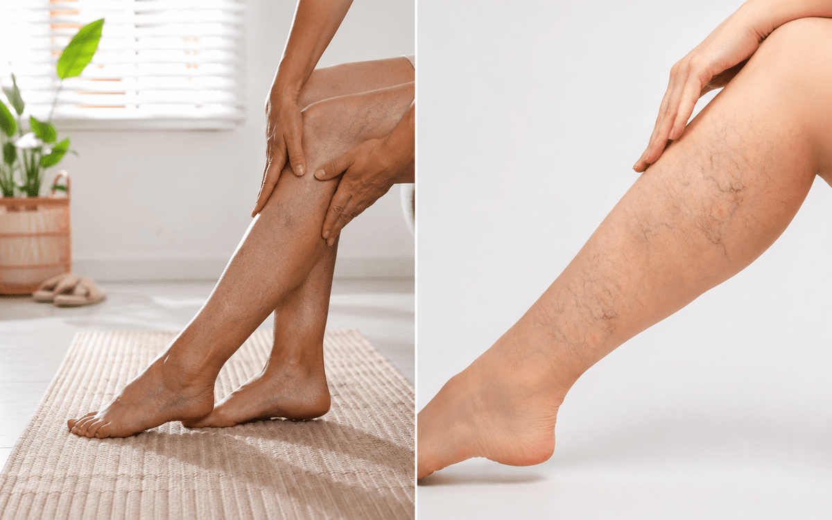
Varicose veins are enlarged, twisted, rope-like veins that bulge visibly beneath the surface of the skin. They most commonly appear on the calves, inner thighs, and backs of the knees, though they can develop anywhere in the lower leg. They are typically blue, dark purple, or flesh-colored and range from a few millimeters to several centimeters in width.
Varicose veins are not simply a cosmetic problem. They are a symptom of chronic venous insufficiency, a condition in which the one-way valves inside the leg veins fail to keep blood moving efficiently toward the heart. When those valves malfunction, blood flows backward and pools in the lower legs. That pooled blood creates sustained pressure inside the vessel walls, causing them to stretch and bulge outward.
According to the National Heart, Lung, and Blood Institute, varicose veins affect a significant portion of adults and are caused by weakened or damaged vein valves that allow blood to collect rather than circulate properly. The condition is progressive. Without treatment, the pressure continues to build, the affected veins continue to enlarge, and the surrounding tissue begins to bear the consequences of sustained circulatory stress.
Common symptoms associated with varicose veins include:
- Aching, throbbing, or heaviness in the legs, particularly after standing or sitting for extended periods
- Swelling in the ankles or lower legs that worsens throughout the day
- Nighttime leg cramps or restless legs
- Itching or burning sensation around the affected veins
- Skin discoloration or hardening near the ankles in more advanced cases
3. What Spider Veins Are
Spider veins are small, thin veins that appear close to the surface of the skin in a web-like or branching pattern. They are typically red, blue, or purple and measure only one to three millimeters in diameter. Unlike varicose veins, they do not bulge above the skin surface. They lie flat and are visible only because the thin overlying skin allows them to be seen.
Spider veins are known medically as telangiectasias. They develop when small blood vessels near the skin dilate and become permanently visible. They appear most commonly on the legs and face, and they affect a significantly larger proportion of the adult population than varicose veins do.
In many cases, spider veins are purely cosmetic and cause no physical symptoms. However, they are not always benign. Spider veins on the legs can sometimes be a surface sign of underlying venous insufficiency, particularly when they appear in clusters, when they are accompanied by symptoms like leg heaviness or aching, or when they are located near areas where deeper vein dysfunction is known to occur.
According to the National Institutes of Health, spider veins and varicose veins share common risk factors and can coexist in the same patient, with spider veins sometimes developing as a result of the same valve dysfunction that produces varicose veins. This is why a medical evaluation that includes duplex ultrasound is the only reliable way to determine whether spider veins are connected to a deeper circulatory problem.
4. Varicose Veins vs Spider Veins: Key Differences
Here is how the two conditions compare across the most important clinical and practical dimensions:
Appearance
- Varicose veins are large, bulging, twisted, and rope-like. They are blue, dark purple, or flesh-colored and raise above the skin surface.
- Spider veins are small, flat, web-like or branching, and red, blue, or purple. They do not bulge above the skin.
Size
- Varicose veins are typically several millimeters to several centimeters wide.
- Spider veins are one to three millimeters wide.
Location
- Varicose veins appear most commonly on the calves, inner legs, and backs of the knees.
- Spider veins appear on the legs and face, often in clusters near the ankles, thighs, or cheeks.
Symptoms
- Varicose veins frequently cause physical symptoms including pain, swelling, heaviness, and cramping.
- Spider veins are often asymptomatic, though they can cause mild aching or burning in some patients.
Underlying cause
- Varicose veins are almost always caused by chronic venous insufficiency and valve dysfunction.
- Spider veins may be caused by valve dysfunction, hormonal changes, sun damage, or simple dilation of small vessels without deeper disease.
Medical significance
- Varicose veins are a progressive medical condition that worsens without treatment and can lead to serious complications. For a full breakdown of what those complications look like over time, our post on what happens when varicose veins go untreated covers each stage in detail.
- Spider veins may be cosmetic only, or may indicate underlying venous disease that warrants evaluation.
5. What Causes Each Type
Both varicose veins and spider veins share several common risk factors, which is why they frequently appear together in the same patient.
Shared risk factors include:
- Family history of vein disease — genetics play a significant role in valve strength and vessel wall integrity
- Age — vein walls and valves weaken naturally over time, with risk increasing significantly after age 40
- Prolonged standing or sitting — occupations that require extended periods on your feet or at a desk increase venous pressure in the lower legs
- Pregnancy — increased blood volume and uterine pressure during pregnancy strain the venous system, and the hormonal changes of pregnancy affect vessel wall tone
- Obesity — excess body weight increases abdominal pressure, which reduces venous return from the lower limbs
- Prior blood clots — deep vein thrombosis can permanently damage vein valves, accelerating the development of both varicose and spider veins
- Hormonal changes — estrogen fluctuations from pregnancy, birth control use, or hormone therapy affect vascular tone and can contribute to spider vein development
What distinguishes their causes:
Varicose veins are specifically caused by valve failure within the larger superficial veins of the leg. The Great Saphenous Vein and Small Saphenous Vein are the most commonly affected vessels. When valves in these veins fail, the resulting reflux creates the pressure and pooling that produces varicose veins.
Spider veins can develop from the same valve dysfunction, but they can also develop independently from local dilation of small superficial vessels without any deeper venous disease. This is why some patients have spider veins without any varicose veins and without any underlying insufficiency.
6. Which One Requires Medical Treatment
This is the question most patients actually want answered, and the honest answer is that it depends on what is driving them.
Varicose veins almost always warrant evaluation. They are a sign of valve dysfunction and chronic venous insufficiency, both of which are progressive. Waiting to see if they improve on their own is not an effective strategy because the underlying cause does not self-resolve. The earlier varicose veins are evaluated and treated, the simpler the treatment and the more complete the outcome.
Spider veins require evaluation when:
- They are accompanied by symptoms such as leg aching, heaviness, swelling, or nighttime cramping
- They are appearing in large numbers or spreading rapidly
- They are located in areas where deeper vein dysfunction is common, particularly around the inner ankle, the back of the knee, or the inner thigh
- You have known risk factors for venous insufficiency including a family history of vein disease, prior blood clots, or a standing occupation
Spider veins may not require medical treatment when:
- They are isolated, stable, and entirely asymptomatic
- They appear on the face and are related to sun damage or rosacea rather than venous disease
- A duplex ultrasound evaluation has confirmed no underlying venous insufficiency
The only reliable way to determine whether your spider veins are connected to a deeper problem is a vein evaluation that includes duplex ultrasound imaging. A visual examination alone cannot distinguish between spider veins caused by superficial vessel dilation and spider veins that are the surface expression of deeper valve dysfunction.
7. Treatment Options for Each Type
Treatment approaches differ between varicose veins and spider veins, though there is some overlap in the methods used.
For varicose veins:
- Radiofrequency ablation (RFA) — the primary treatment for varicose veins caused by insufficiency in the Great or Small Saphenous Vein. A catheter delivers controlled radiofrequency energy to collapse and seal the malfunctioning vein from the inside. Blood is naturally rerouted through healthier vessels.
- Endovenous laser ablation (EVLA) — similar to RFA but uses laser energy rather than radiofrequency. Effective for certain vein anatomies where RFA may be less suitable.
- Ambulatory phlebectomy — removes surface-level varicose veins through small micro-incisions, often performed alongside ablation procedures to address bulging veins not accessible by catheter.
- VenaSeal — uses medical-grade adhesive to close the vein without thermal energy, suitable for patients who prefer a non-heat-based approach.
For spider veins:
- Sclerotherapy — the most effective and widely used treatment for spider veins. A specialized solution is injected directly into the affected vessels, causing them to collapse, fade, and be reabsorbed by the body over several weeks. Multiple sessions are often required depending on the extent of the veins.
- Surface laser treatment — used for very small spider veins, particularly on the face, where injection-based treatment is less practical.
When spider veins are caused by underlying venous insufficiency, treating the surface veins alone without addressing the deeper valve problem typically produces poor long-term results. The spider veins are likely to return. In these cases, the appropriate sequence is to treat the underlying insufficiency first, then address any remaining surface veins with sclerotherapy.
For a complete overview of all vein treatment options available at Advanced Medical and Diagnostics Center, visit our vein treatment service page.
8. Frequently Asked Questions
Spider veins themselves do not transform into varicose veins, but they can coexist with the same underlying venous insufficiency that produces varicose veins. If you develop spider veins and notice varicose veins appearing alongside them, or if your spider veins are accompanied by leg symptoms, a duplex ultrasound evaluation is warranted to assess whether deeper valve dysfunction is present.
Varicose veins are a sign of chronic venous insufficiency, which is a progressive condition. Without treatment, they can lead to serious complications including skin changes, venous ulcers, and superficial blood clots. They are not typically life-threatening in their early stages, but the risks associated with leaving them untreated increase significantly over time.
Over-the-counter creams and topical treatments do not treat spider veins. They may temporarily improve the appearance of surrounding skin but do not close or remove the affected vessels. Medical treatment through sclerotherapy or laser therapy is required to eliminate spider veins. A physician evaluation first ensures that any underlying venous disease is identified before surface treatment begins.
You cannot prevent vein disease entirely if you have a genetic predisposition, but you can reduce your risk and slow progression. Regular walking, maintaining a healthy body weight, avoiding prolonged standing or sitting without movement breaks, wearing compression stockings in high-risk occupations, and elevating your legs when resting all support healthy venous circulation.
A visual assessment gives you a starting point. Bulging, rope-like, twisted veins that raise above the skin surface are varicose veins. Flat, web-like, small veins in red, blue, or purple are spider veins. Many patients have both. A duplex ultrasound performed by a vein specialist is the definitive diagnostic tool that identifies which veins have valve dysfunction, how severe the insufficiency is, and which treatment approach is most appropriate.
9. What to Do If You Are Unsure Which Type You Have
Identifying whether you have varicose veins, spider veins, or both is a starting point, not a diagnosis. The appearance of veins on the surface tells you something is happening, but it does not tell you what is driving it or how far the underlying disease has progressed.
If you have been noticing veins on your legs and are unsure whether they warrant evaluation, the answer in most cases is yes. A vein evaluation is a non-invasive, straightforward process that gives you a clear picture of your venous health. It identifies whether underlying insufficiency is present, how advanced it is, and what treatment approach is most appropriate for your specific situation.
Our teams at our Hackensack, NJ office and our Wayne, NJ office provide comprehensive vein evaluations including duplex ultrasound for patients throughout New Jersey. For full details on what vein treatment at our Hackensack location involves, visit our vein treatment in Hackensack, NJ page.
Key Takeaways
- Varicose veins are large, bulging, twisted veins caused by chronic venous insufficiency and valve failure — they are a progressive medical condition
- Spider veins are small, flat, web-like vessels that may be cosmetic only or may signal underlying venous disease
- The key difference between varicose veins vs spider veins is size, appearance, symptoms, and medical significance
- Both share common risk factors including genetics, age, prolonged standing, pregnancy, and obesity
- Varicose veins almost always warrant evaluation and treatment — spider veins warrant evaluation when symptomatic, spreading, or accompanied by risk factors
- Varicose veins are treated with radiofrequency ablation, laser ablation, VenaSeal, or phlebectomy — spider veins are treated with sclerotherapy or surface laser
- A duplex ultrasound is the only reliable way to determine whether spider veins are connected to deeper venous disease
WHAT YOU SEE ON THE SURFACE IS NOT ALWAYS THE COMPLETE PICTURE OF WHAT IS HAPPENING IN YOUR VEINS.
A duplex ultrasound is the only reliable way to assess venous valve function and determine whether the veins you are seeing are purely cosmetic or connected to an underlying circulatory condition that warrants treatment. Speaking with a physician who can evaluate both the surface presentation and the deeper venous anatomy gives you the complete picture you need to make an informed decision.
Disclaimer: The information in this article is for educational purposes only and does not constitute medical advice, diagnosis, or treatment recommendations. All decisions regarding vein evaluation and treatment should be made in consultation with a board-certified physician who is familiar with your complete medical history, current symptoms, and individual health circumstances.