Advanced Medical & Diagnostic Center

man experiencing early signs of lung disease

What Are the Early Signs of Lung Disease That Most People Ignore?

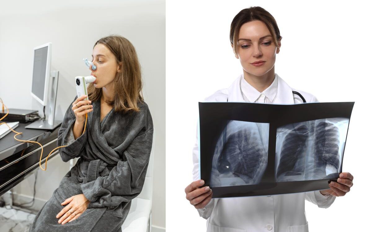
What Are the Early Signs of Lung Disease That Most People Ignore? May 24, 2026 10 min read Table of Contents 1. What Are the Early Signs of Lung Disease and Why Are They So Easy to Miss? 2. Why First Responders Face Higher Risk for Early Lung Disease 3. Conditions Associated With Early Lung Disease Signs 4. How Early Lung Disease Is Detected Through Testing 5. When You Should Not Wait to Get Evaluated 6. What to Expect From Your Pulmonary Evaluation 7. Frequently Asked Questions 8. The Bottom Line on Early Signs of Lung Disease Most of us do not think about lung health until something feels noticeably wrong. You write off a persistent cough as allergies. You attribute shortness of breath after climbing stairs to being out of shape. You dismiss feeling winded during a workout that used to feel easy as just a bad day. But these are often the early signs of lung disease, and dismissing them is exactly how lung conditions progress undetected for years before becoming serious. Understanding the early signs of lung disease and knowing when they warrant a medical evaluation can make a significant difference in your long-term respiratory health. This is especially true if you are a first responder, a smoker, or someone who has worked in environments with regular exposure to smoke, chemicals, or airborne particles. 1. What Are the Early Signs of Lung Disease and Why Are They So Easy to Miss? The reason early signs of lung disease are so frequently ignored is that they develop gradually and mimic conditions most people consider minor or normal. Unlike a broken bone or an acute infection, lung disease in its early stages rarely announces itself with unmistakable pain or sudden onset. It creeps in slowly, and by the time your symptoms become impossible to ignore, the underlying condition has often been developing for years. According to the American Lung Association, many people do not seek medical evaluation for respiratory symptoms until their lung function has already declined significantly. This delay in diagnosis is one of the primary reasons lung disease is often more advanced by the time it is identified. The most commonly ignored early signs of lung disease include: A cough that has lasted more than three weeks — if your cough does not resolve after a respiratory infection or has been present for an extended period without a clear cause, this is one of the most consistent early indicators of underlying lung disease Shortness of breath during activities that previously felt manageable — if you are getting winded on stairs, during moderate exercise, or during tasks that did not used to cause breathlessness, that is a meaningful change worth investigating Wheezing or a whistling sound when you breathe — this indicates airway narrowing or obstruction and is associated with both asthma and early COPD Tightness or pressure in your chest — not always cardiac in origin, chest tightness can reflect airway inflammation or reduced lung compliance Increased mucus production — if you are producing more mucus than usual, particularly in the morning, this can indicate chronic bronchitis or other inflammatory lung conditions Frequent respiratory infections — if your lungs are not functioning optimally, you become more susceptible to infections, and a pattern of repeated respiratory illness can signal underlying disease Fatigue during physical activity — when your lungs are not efficiently exchanging oxygen and carbon dioxide, your body works harder to compensate, producing fatigue that feels disproportionate to the level of exertion 2. Why First Responders Face Higher Risk for Early Lung Disease If you are a police officer, firefighter, or EMS worker, the early signs of lung disease carry additional significance because of the occupational exposures that come with your job. According to the National Institute for Occupational Safety and Health, firefighters face significantly elevated risk of respiratory disease compared to the general population due to cumulative exposure to combustion byproducts, particulate matter, and toxic chemicals encountered at fire scenes. Even with proper protective equipment, repeated exposure over your career contributes to measurable changes in lung function. Specific occupational factors that increase your lung disease risk as a first responder include: Smoke inhalation at fire scenes, including structural fires where burning synthetic materials release particularly toxic compounds Exposure to diesel exhaust in fire stations and other enclosed environments Chemical exposures during hazmat incidents or industrial emergencies Particulate matter exposure during vehicle accidents, building collapses, or construction-related emergencies Repeated use of respiratory protection equipment that, while essential, does not eliminate all exposure risk Many firefighters and first responders develop early signs of lung disease well before retirement age, and those changes are often first detected during pulmonary function testing rather than through your own symptom recognition. This is why routine respiratory screening is a standard recommendation for your population. 3. Conditions Associated With Early Lung Disease Signs Understanding which conditions produce the early signs you might be ignoring helps clarify why prompt evaluation matters. The most common underlying conditions associated with these symptoms include: Chronic Obstructive Pulmonary Disease (COPD) COPD is an umbrella term for progressive lung conditions including chronic bronchitis and emphysema. According to the Global Initiative for Chronic Obstructive Lung Disease, COPD affects hundreds of millions of people globally and is significantly underdiagnosed because your early symptoms are mild and easy to dismiss. The earlier your COPD is identified, the more effectively its progression can be managed. Asthma Adult-onset asthma is more common than most people realize, and it does not always present with the dramatic wheezing episodes you may associate with the condition. Mild persistent asthma can produce subtle symptoms including slight breathlessness during exertion and occasional chest tightness that you might attribute to stress or your fitness level. Occupational Lung Disease Occupational lung diseases including hypersensitivity pneumonitis, occupational asthma, and pneumoconiosis develop from repeated workplace exposures. These conditions often begin with symptoms that are easy for you to attribute to other causes, and they progress more rapidly with continued exposure. Pulmonary Fibrosis

doctor explaining about asymptomatic heart disease

Can You Have a Heart Problem With No Symptoms at All?

Can You Have a Heart Problem With No Symptoms at All? If you have a serious heart problem, you will probably feel it. Chest pain, shortness of breath, dizziness and these are the warning signs you have likely been taught to watch for. But asymptomatic heart disease is far more common than most people realize, and it is one of the primary reasons that cardiovascular screening exists in the first place. The absence of symptoms does not mean the absence of disease. If you are a man over 35, a first responder, or someone with known risk factors, understanding what asymptomatic heart disease is and how it is detected can be the difference between catching a problem early and facing a cardiac event with no prior warning. What Is Asymptomatic Heart Disease and How Common Is It? Asymptomatic heart disease refers to cardiac conditions that are present and progressing without producing noticeable symptoms. The most common form is atherosclerosis, which is the gradual buildup of plaque inside the arterial walls. This process can develop over decades without causing pain, discomfort, or any sensation that something is wrong. According to the American Heart Association, approximately half of all cardiac events, including heart attacks, occur in people who had no prior symptoms or diagnosis of heart disease. Many of these individuals had risk factors that, if identified and addressed earlier, could have reduced or prevented the event entirely. Asymptomatic heart disease is particularly relevant for: Men over the age of 35, particularly those with a family history of heart disease First responders including police officers, firefighters, and EMS workers who face chronic occupational stress and physical exertion Men with elevated blood pressure, high cholesterol, or elevated blood sugar who have not yet experienced symptoms Individuals who smoke or have smoked in the past Men who are overweight or have significant abdominal fat People with sedentary lifestyles or disrupted sleep patterns, including shift workers The challenge with asymptomatic heart disease is that by the time symptoms appear, the underlying condition has often been developing for years. Why First Responders Face Higher Risk for Undetected Cardiac Problems Police officers, firefighters, and EMS workers are at elevated risk for cardiovascular disease compared to the general population, and a significant portion of that risk is asymptomatic until a critical event occurs. According to the National Institute for Occupational Safety and Health, sudden cardiac death accounts for a significant proportion of line-of-duty deaths among firefighters, and many of these events involve underlying coronary artery disease that was not previously identified. The physical and psychological demands of first responder work place sustained stress on the cardiovascular system in ways that accelerate the progression of conditions that may not produce symptoms for years. Contributing factors specific to first responder cardiovascular risk include: Repeated exposure to high-intensity physical exertion during emergency responses, which places acute demand on a heart that may have underlying disease Chronic occupational stress that elevates cortisol and inflammatory markers over time Disrupted sleep from shift work, which is independently associated with elevated cardiovascular risk Exposure to smoke, chemicals, and environmental toxins that affect arterial health over a career Deferred medical care, as many first responders avoid seeking evaluation unless symptoms are severe The combination of these factors makes routine cardiovascular screening particularly important for this population, even in the absence of any symptoms. How Asymptomatic Heart Disease Is Detected Because asymptomatic heart disease produces no warning signs by definition, the only reliable way to detect it is through diagnostic testing. This is exactly what cardiovascular screening is designed to do. According to the Centers for Disease Control and Prevention, routine cardiovascular screening can identify risk factors and early-stage disease before a cardiac event occurs, giving patients and providers the opportunity to intervene with lifestyle changes, medication, or closer monitoring. Common tests used to detect asymptomatic heart disease include: Electrocardiogram (EKG) — measures the electrical activity of the heart and can identify irregular rhythms, prior silent heart attacks, and structural abnormalities Stress test — evaluates how the heart responds to physical exertion and can reveal blood flow problems that are not apparent at rest Echocardiogram — uses ultrasound to assess heart structure, valve function, and wall motion, identifying abnormalities that produce no symptoms Blood pressure monitoring — consistently elevated blood pressure is one of the most common and underrecognized contributors to asymptomatic cardiac damage Lipid panel — high LDL cholesterol and elevated triglycerides contribute directly to plaque buildup in arterial walls without causing any noticeable symptoms Blood glucose and metabolic markers — elevated blood sugar and insulin resistance accelerate atherosclerosis and increase cardiac risk For a detailed overview of what a comprehensive evaluation involves, our guide on how cardiovascular and pulmonary testing works covers each test and what it measures. The Silent Heart Attack: When Asymptomatic Heart Disease Progresses Undetected One of the most striking manifestations of asymptomatic heart disease is the silent heart attack, a myocardial infarction that occurs without the classic symptoms of chest pain, arm pain, or shortness of breath. Many silent heart attacks are only discovered during a subsequent EKG or imaging study. According to the American College of Cardiology, silent myocardial infarctions account for a meaningful percentage of all heart attacks and are associated with significant long-term cardiovascular risk if not identified and managed. Men are more likely than women to experience a classic symptomatic heart attack, but silent events occur across both sexes and all age groups. For first responders and physically active men, a silent heart attack is particularly dangerous because the individual may continue performing at a high physical level without knowing that cardiac damage has occurred. Each subsequent high-intensity exertion places additional demand on an already compromised heart. This is one of the strongest arguments for routine screening even in men who feel completely healthy. An EKG performed as part of a standard cardiovascular evaluation can reveal evidence of a prior silent event that the patient had no awareness of. What Happens After Asymptomatic Heart Disease Is

Alcohol effect on testosterone levels in men

How Does Alcohol Affect Testosterone Levels in Men?

How Does Alcohol Affect Testosterone Levels in Men? If you have been experiencing fatigue, low drive, or difficulty building muscle and you also drink regularly, you may be wondering whether there is a connection. The alcohol effect on testosterone is a well-documented area of research, and the findings are relevant for any man who is either concerned about low testosterone or currently undergoing testosterone replacement therapy. This article explains what the research shows, how much alcohol it takes to affect hormone levels, and what men who are on TRT or considering it should know about drinking and their hormonal health. What the Research Says About Alcohol Effect on Testosterone The relationship between alcohol consumption and testosterone is not a simple one. A single drink at a social event is not going to crash your hormone levels. But consistent, heavy drinking has a measurable and clinically significant impact on testosterone production that every man should understand. According to the National Institute on Alcohol Abuse and Alcoholism, alcohol interferes with the function of the hypothalamic-pituitary-gonadal axis, which is the hormonal signaling chain responsible for regulating testosterone production. When alcohol disrupts this system, the result is reduced testosterone output at multiple levels. The specific mechanisms through which alcohol affects testosterone include: Direct toxic effects on the Leydig cells in the testes, which are responsible for producing testosterone Increased conversion of testosterone to estrogen through a process called aromatization, which is accelerated by alcohol metabolism in the liver Elevated cortisol levels caused by alcohol consumption, and cortisol directly suppresses testosterone production Disruption of sleep architecture, particularly REM sleep, during which a significant portion of daily testosterone production occurs Impaired liver function in heavy drinkers, which affects how the body processes and clears hormones Each of these mechanisms operates independently, which means heavy drinking hits testosterone from multiple directions at once. How Much Alcohol Actually Lowers Testosterone? This is the question most men actually want answered, and the honest answer is that it depends on frequency, quantity, and individual physiology. According to a study published in the Journal of Clinical Endocrinology and Metabolism, acute alcohol intoxication in healthy men produced a significant reduction in serum testosterone levels within hours of consumption. The effect was dose-dependent, meaning larger amounts produced greater reductions. For context, here is what the research generally shows across drinking patterns: Occasional light drinking — one to two drinks a few times per week produces minimal long-term impact on baseline testosterone levels in most healthy men Moderate daily drinking — two to three drinks per day consistently begins to suppress testosterone production over time, particularly when combined with other lifestyle factors like poor sleep or high stress Heavy or binge drinking — four or more drinks in a session, or consistent heavy daily consumption produces clinically meaningful reductions in testosterone and can contribute to symptoms of hypogonadism Chronic alcoholism — long-term heavy alcohol dependence is associated with severe testosterone suppression, testicular atrophy, and significantly elevated estrogen levels in men The alcohol effect on testosterone is cumulative. A single heavy night may cause a temporary dip that recovers within a few days. Consistent heavy drinking over months or years creates a sustained suppression that does not resolve simply by cutting back. Alcohol, Estrogen, and the Hormonal Imbalance Men Do Not Expect One aspect of the alcohol effect on testosterone that surprises many men is the estrogen connection. Alcohol does not just lower testosterone. It also raises estrogen levels in men, and the combination of the two creates a hormonal imbalance that amplifies the symptoms most men associate with low T. According to the National Institutes of Health, alcohol consumption increases the activity of aromatase, an enzyme that converts testosterone into estradiol, which is a form of estrogen. In men, elevated estrogen levels contribute to: Increased body fat, particularly around the chest and midsection Reduced muscle mass and strength Mood changes including irritability and emotional flatness Reduced libido and sexual function Water retention and bloating These symptoms overlap heavily with the symptoms of low testosterone. For men who drink regularly and are experiencing these issues, the alcohol effect on testosterone and estrogen simultaneously may be a significant contributing factor. You can review a full breakdown of low testosterone symptoms and how to recognize them to better understand how the two conditions overlap. What Men on TRT Should Know About Alcohol If you are currently on testosterone replacement therapy, the alcohol effect on testosterone takes on added significance. TRT works by supplementing the testosterone your body is not producing at adequate levels. Alcohol does not cancel out TRT directly, but it does create several complications worth understanding. Alcohol can blunt the effectiveness of TRT. If you are drinking heavily while on therapy, the elevated cortisol and aromatase activity caused by alcohol will work against the goals of your treatment. Your provider may find it harder to optimize your levels, and you may not experience the full benefits of the protocol. Alcohol affects the cardiovascular markers your provider monitors. TRT protocols for first responders and other patients include regular monitoring of hematocrit, blood pressure, and lipid panels. Heavy alcohol consumption raises blood pressure, elevates triglycerides, and affects liver enzymes, all of which appear in your follow-up labs. These changes can complicate your provider’s ability to manage your protocol safely. Sleep disruption compounds the hormonal impact. Alcohol is widely understood to interfere with sleep quality, and a significant portion of daily testosterone production occurs during deep sleep. Men on TRT who drink heavily may find their morning testosterone levels are inconsistent, making it harder to assess whether the protocol is working as intended. Liver health matters for hormone processing. The liver plays a central role in metabolizing hormones, including testosterone. Heavy alcohol use impairs liver function over time, which can affect how your body processes and clears the testosterone introduced through TRT. According to the American Liver Foundation, chronic alcohol use is one of the leading causes of liver damage, and compromised liver function has direct implications

First responder in TRT fitness for duty clearance

Can TRT Affect Your Fitness for Duty Clearance as a Police Officer or Firefighter?

Can TRT Affect Your Fitness for Duty Clearance as a Police Officer or Firefighter? If you are a police officer, firefighter, or EMS worker considering testosterone replacement therapy, one of the first questions you probably have is whether TRT fitness for duty status will be affected. It is a fair concern. Your job depends on passing medical evaluations, and the last thing you want is a treatment that raises questions during your next department physical or clearance review. The short answer is that TRT does not automatically disqualify you from duty. But the details matter, and understanding how your department evaluates hormone therapy can help you make informed decisions about your health without putting your career at risk. This guide covers what first responders need to know before, during, and after starting TRT. What Is a Fitness for Duty Evaluation and Who Requires One? A fitness for duty evaluation is a medical assessment used by law enforcement agencies, fire departments, and EMS organizations to determine whether an employee is physically and mentally capable of performing the essential functions of their job. These evaluations are typically required at hiring, on a periodic basis, after an injury or illness, or when a supervisor has concerns about an employee’s capacity to perform safely. For firefighters, the most widely referenced standard is NFPA 1582, which is the National Fire Protection Association’s guide for occupational medical evaluations. For law enforcement, standards vary by department and state but generally follow guidelines from the International Association of Chiefs of Police or equivalent state-level frameworks. Neither NFPA 1582 nor most law enforcement medical guidelines automatically disqualify a candidate or active employee for being on testosterone replacement therapy. What matters to evaluating physicians is whether the condition being treated, and the treatment itself, affects your ability to safely perform job functions. Does TRT Show Up in Department Physicals or Drug Screenings? This is one of the most common questions first responders have about TRT fitness for duty evaluations, and it deserves a direct answer. Standard occupational drug screenings used by most police and fire departments test for controlled substances including opioids, stimulants, benzodiazepines, and cannabinoids. Testosterone is not included on standard SAMHSA-5 or DOT drug panels. According to the Substance Abuse and Mental Health Services Administration, federally mandated workplace drug testing panels do not include testosterone or other hormone replacement therapies. However, some departments conduct separate medical evaluations that include hormone panels as part of a broader health assessment. If your department orders bloodwork as part of your physical, testosterone levels may be reviewed. In that context, being on a medically supervised TRT protocol with documented lab values is generally viewed favorably, because it demonstrates you are under professional medical care and being monitored consistently. The key distinction is between a drug screening and a medical evaluation. TRT will not flag a drug test. It may appear in a medical panel, and in that context, documentation from your provider is your best protection. How TRT Fitness for Duty Is Evaluated by Occupational Physicians When an occupational physician reviews a first responder who is on testosterone replacement therapy, they are not simply looking at whether the person takes medication. They are evaluating functional capacity. According to the American College of Occupational and Environmental Medicine, occupational fitness evaluations focus on functional ability rather than the presence of a specific medical condition or treatment. The key questions an occupational physician is asking during a TRT fitness for duty review are: Can this person perform the physical demands of the job safely? Are their cardiovascular markers within acceptable ranges? Is their testosterone treatment stable and properly supervised? Are lab values documented and within therapeutic range? Is the patient being monitored consistently by a qualified provider? A first responder on a well-managed TRT protocol, with stable hormone levels and normal cardiovascular markers, is generally in a strong position during a fitness for duty review. It is also worth understanding what creates problems during these evaluations. The risk is not the treatment itself. It is untreated low testosterone. A man with significantly low testosterone levels may show declines in: Physical performance and strength Cognitive sharpness and reaction time Stress tolerance and mood stability Energy levels during prolonged physical exertion All of these are directly relevant to job performance in law enforcement and fire service. Addressing that deficiency with supervised TRT often improves the very metrics that fitness evaluations are designed to measure. For this reason, many occupational health physicians who work with fire and law enforcement departments view appropriately managed TRT positively when labs are in range and the patient is being monitored regularly. What First Responders Should Know Before Starting TRT If you are a police officer, firefighter, or EMS worker considering TRT, taking the right steps before starting treatment protects both your health and your career. Get a full diagnostic workup first. Do not start testosterone therapy based on symptoms alone. You need documented lab results confirming low testosterone, along with a full panel that includes cardiovascular markers, red blood cell counts, and prostate-specific antigen levels. This documentation is important if your treatment is ever reviewed during a fitness evaluation. Understanding what a testosterone replacement therapy program involves and what the evaluation process looks like can help you prepare. Work with a provider experienced in occupational health. A physician who understands the physical demands placed on first responders can tailor your protocol to support your job performance. Not every TRT provider has experience with this patient population, so it is worth asking whether your doctor is familiar with department medical standards and fitness for duty requirements. Keep complete records. Every lab result, every dosage adjustment, and every follow-up visit should be documented. If your department ever requests medical records related to a fitness for duty review, having a clean and complete file from your treating physician makes the process significantly smoother. Understand your department’s specific policies. Medical standards vary across departments, counties, and states. Some departments have specific disclosure requirements for ongoing

woman experiencing effects of night shifts on health

How Years of Night Shifts Affect the Heart and Lungs of First Responders

How Years of Night Shifts Affect the Heart and Lungs of First Responders Most people understand that being a first responder is physically and mentally demanding. What is less understood is how the structure of the work itself, specifically years of night shifts, rotating schedules, and sleep disruption, quietly damages the cardiovascular and respiratory systems over time. The effects of night shifts on health are cumulative, often invisible for years, and frequently misattributed to aging or stress rather than identified as a preventable occupational health risk. For police officers, firefighters, and EMTs who have spent years working overnight rotations or 24-hour tours, understanding what those shifts are doing to the heart and lungs is not just useful information. It is clinically important. The conditions that develop from chronic shift work are serious, they are largely silent until they become acute, and they are far more common in first responders than in the general population. This blog explains exactly how night shift work affects cardiovascular and pulmonary health, what the warning signs look like, and what first responders can do to protect themselves before symptoms appear. Why Night Shifts Are Harder on the Body Than Day Work The human body is governed by a 24-hour internal clock called the circadian rhythm. This biological system regulates sleep and wake cycles, hormone release, body temperature, metabolism, immune function, and cardiovascular activity. Every major organ system in the body including the heart and lungs operates on a schedule set by this internal clock. Night shift work forces the body to function against its natural rhythm. Rather than sleeping when the circadian system is signaling rest and repair, night shift workers are awake, alert, and often physically active. Rather than being active during daylight hours when the body is primed for performance, they are trying to sleep, often poorly and incompletely. According to the National Institutes of Health, chronic circadian rhythm disruption is associated with increased risk of cardiovascular disease, metabolic dysfunction, and immune system impairment. The longer the duration of shift work exposure, the more significant the cumulative health impact. For first responders who may spend an entire career on rotating or overnight schedules, this is not a short-term disruption. It is a decades-long physiological challenge that the body never fully adapts to. The Effects of Night Shifts on Heart Health The cardiovascular system is one of the most vulnerable organ systems to the effects of night shifts on health. Multiple mechanisms connect shift work to increased cardiac risk, and they tend to compound each other over time. Elevated Blood Pressure Blood pressure follows a natural daily pattern, rising in the morning and dipping during nighttime sleep. This overnight dip is called nocturnal dipping and it plays an important role in cardiovascular recovery and repair. Night shift workers who sleep during the day often lose this nocturnal dip entirely, meaning their blood pressure never gets the regular recovery period it needs. According to the American Heart Association, non-dipping blood pressure patterns are associated with significantly higher risk of heart attack, stroke, and left ventricular hypertrophy compared to normal dipping patterns. Over years of night shift work, this persistent elevation contributes to chronic hypertension and accelerated cardiovascular aging. Increased Inflammation Sleep deprivation triggers the release of pro-inflammatory cytokines, proteins that signal the immune system to respond as if the body is under threat. Chronic low-grade inflammation is one of the primary drivers of atherosclerosis, the buildup of plaque inside arterial walls that leads to heart attack and stroke. First responders who work night shifts for years accumulate a significant inflammatory burden that standard annual physicals often do not measure or address. Disrupted Cholesterol and Metabolic Function Night shift workers consistently show less favorable cholesterol profiles compared to day workers, with higher levels of LDL cholesterol and triglycerides and lower levels of protective HDL cholesterol. Insulin sensitivity also decreases with chronic sleep disruption, raising the risk of type 2 diabetes, which is itself a major cardiovascular risk factor. Higher Risk of Cardiac Events on Duty Heart attack is one of the leading causes of line of duty death among firefighters and police officers. Research has consistently shown that cardiac events during or immediately after physically demanding calls are disproportionately common in first responders, and that underlying cardiovascular disease accumulated through years of shift work is a major contributing factor. The exertion of emergency response places a sudden acute demand on a cardiovascular system that has been silently weakened over years. The Effects of Night Shifts on Lung Health The respiratory system is affected by shift work through several overlapping mechanisms, and the effects are compounded for first responders who also face direct occupational pulmonary hazards. Reduced Immune Defense in the Airways The lungs rely on a well-functioning mucociliary clearance system to trap and expel inhaled particles, pathogens, and irritants. This system functions most efficiently during periods of adequate rest. Chronic sleep deprivation impairs mucociliary function, leaving the airways more vulnerable to infection, irritation, and long-term damage from inhaled hazards. For firefighters and other first responders exposed to smoke, dust, and chemical fumes, this reduced airway defense means that the lungs are less equipped to handle occupational exposures when sleep is chronically disrupted. Increased Risk of Respiratory Infections Sleep deprivation is one of the most well-documented risk factors for susceptibility to respiratory infections. First responders working night shifts are more likely to develop upper and lower respiratory infections, and those infections tend to be more severe and longer lasting than in well-rested individuals. Over years, repeated respiratory infections contribute to cumulative airway damage and increased risk of chronic respiratory conditions. Accelerated Decline in Lung Function Lung function naturally declines with age, but the rate of decline is significantly accelerated by chronic sleep deprivation, inflammation, and repeated occupational exposure without adequate recovery. First responders who have spent careers on night shifts and who have also experienced regular smoke or chemical exposure face a compounded rate of lung function decline that may not become symptomatic until significant capacity

Pulmonary Function Test vs Chest X-Ray Comparison

Pulmonary Function Test vs Chest X-Ray: What Is the Difference and Which One Do You Need?

Pulmonary Function Test vs Chest X-Ray: What Is the Difference and Which One Do You Need? If your doctor has recommended lung testing, you may be wondering what the difference is between a pulmonary function test vs chest x-ray and which one is actually right for your situation. Both are used to evaluate respiratory health, but they measure completely different things, detect different conditions, and serve different clinical purposes. Knowing the difference helps you understand what your provider is looking for and why one test may be recommended over the other, or why both might be needed at the same time. This blog explains what each test does, what it can and cannot detect, how they compare directly, and which situations call for which approach. If you want a deeper look at how a PFT works on its own before reading the comparison, our guide on what a pulmonary function test is and what it measures is a helpful starting point. What Is a Pulmonary Function Test? A pulmonary function test, commonly called a PFT, is a non-invasive breathing test that measures how well your lungs work. Rather than producing an image of the lungs, a PFT measures the functional performance of your respiratory system, how much air you can inhale and exhale, how fast you can move air in and out, and how efficiently your lungs transfer oxygen into the bloodstream. The most common component of a pulmonary function test is spirometry, which measures: Forced vital capacity (FVC) – the total amount of air you can exhale after a full breath Forced expiratory volume in one second (FEV1) – how much air you can push out in the first second of exhalation The FEV1/FVC ratio – a key indicator used to identify airflow obstruction Additional PFT measurements may include lung volume testing, which assesses total lung capacity and residual volume, and diffusion capacity testing, which evaluates how efficiently oxygen crosses from the air sacs in the lungs into the bloodstream. According to the American Thoracic Society, pulmonary function testing is the standard diagnostic tool for detecting, classifying, and monitoring obstructive and restrictive lung diseases including asthma, COPD, and occupational lung conditions. A pulmonary function test is effort-dependent, meaning the accuracy of results depends on the patient breathing as hard and as fully as possible during the test. A trained technician guides you through the process to ensure results are reliable. What Is a Chest X-Ray? A chest x-ray is a form of medical imaging that produces a two-dimensional picture of the structures inside the chest, including the lungs, heart, ribs, and major blood vessels. Unlike a pulmonary function test, a chest x-ray does not measure how the lungs perform. It shows what the lungs look like structurally. A chest x-ray can reveal: Pneumonia or lung infections Fluid around or in the lungs Collapsed lung (pneumothorax) Tumors or masses Enlarged lymph nodes Rib fractures or structural abnormalities Signs of heart enlargement or failure Significant scarring or fibrosis visible on imaging A chest x-ray is quick, widely available, and exposes patients to a small amount of radiation. It is often one of the first imaging tools used when a patient presents with chest pain, shortness of breath, or a suspected infection. According to the National Institutes of Health, chest x-rays are one of the most frequently performed diagnostic imaging studies in medicine, but they have significant limitations when it comes to detecting early or functional lung disease. Pulmonary Function Test vs Chest X-Ray: The Key Differences Understanding how these two tests compare directly is the clearest way to understand when each one is appropriate.   Pulmonary Function Test Chest X-Ray Measures Airflow, lung volume, gas exchange Lung and chest structure appearance Detects Asthma, COPD, airflow obstruction Infections, fluid, masses, abnormalities Misses Tumors, infections, fluid, masses Early functional lung disease Radiation None Small dose of ionizing radiation Results Measurements vs predicted normal values Visual findings by a radiologist Best For Diagnosing and monitoring lung function Ruling out structural problems quickly Duration 30 to 60 minutes 10 to 15 minutes Effort Yes – active breathing required No – patient holds still When a Pulmonary Function Test Is the Right Choice A pulmonary function test is typically the better diagnostic tool when: You have symptoms of asthma, COPD, or chronic bronchitis and a diagnosis has not yet been confirmed You are being evaluated for occupational lung disease related to smoke, dust, chemical, or toxin exposure Your provider needs to assess the severity of a known lung condition and track whether it is progressing or improving You are a first responder, firefighter, or worker with regular exposure to airborne hazards You have unexplained shortness of breath during physical activity with no structural findings on imaging Pre-surgical evaluation is needed to assess whether your lungs can handle anesthesia and recovery Monitoring is required for a lung condition being treated with medication For first responders and workers in physically demanding roles, pulmonary function testing provides a functional baseline that imaging cannot. A firefighter may have a normal chest x-ray and still have measurably reduced lung capacity from years of smoke exposure. A PFT catches that where imaging does not. When a Chest X-Ray Is the Right Choice A chest x-ray is typically the better first step when: You have symptoms of pneumonia such as fever, productive cough, and chest pain A lung infection or fluid accumulation is suspected There is concern about a mass, tumor, or enlarged lymph nodes in the chest You have experienced trauma to the chest and rib fractures or pneumothorax need to be ruled out Shortness of breath is sudden in onset and a structural cause needs to be identified quickly Heart enlargement or pulmonary edema is suspected based on symptoms A chest x-ray answers the question of whether something looks abnormal in the chest. A pulmonary function test answers the question of whether the lungs are functioning normally. These are different questions, and both are valid depending on what the clinical

low testosterone in first responders symptoms

How Low Testosterone in First Responders Affects Police, Fire, and EMS – What You Need to Know About TRT

How Low Testosterone in First Responders Affects Police, Fire, and EMS – What You Need to Know About TRT First responders are trained to push through discomfort. Police officers, firefighters, and EMTs show up regardless of how they feel, perform under pressure, and rarely ask for help. That mindset is part of what makes them effective at their jobs. It is also part of what makes low testosterone in first responders so easy to miss. The symptoms develop gradually. Fatigue that used to go away after a good night of sleep starts becoming chronic. Strength and endurance that once felt natural begin to decline. Mood becomes harder to manage. Most first responders attribute these changes to the job, to age, or to the cumulative weight of years on the front line. What many do not realize is that their career itself may be directly accelerating hormonal decline, and that there is a clinically proven way to address it. This blog covers why low testosterone in first responders is more common than most people realize, how the demands of frontline work affect hormone levels, what the symptoms look like, and what TRT can do about it. If you are not yet familiar with how testosterone replacement therapy works, our guide on what testosterone replacement therapy is and how it works is a helpful place to start before reading further. Why First Responders Are at Higher Risk for Low Testosterone Testosterone production is highly sensitive to the body’s overall stress load. When the body is under chronic physical or psychological stress, it prioritizes survival over hormone production. For most men, this is an occasional response. For first responders, it is a daily reality. Several specific factors unique to frontline careers drive testosterone decline faster than normal aging alone. Shift Work and Disrupted Sleep Testosterone is primarily produced during deep sleep. Men who work rotating shifts, overnight rotations, or extended 24-hour tours consistently disrupt the sleep cycles their bodies depend on for hormone production. According to the National Institutes of Health, sleep deprivation and circadian rhythm disruption are directly associated with reduced testosterone production in men. A firefighter working a 24-hour shift followed by irregular sleep at home is not giving his body the recovery window it needs to maintain healthy hormone levels. Chronic Stress and Elevated Cortisol The relationship between cortisol and testosterone is one of opposition. When cortisol rises, testosterone falls. First responders experience cortisol spikes repeatedly throughout their careers, responding to emergencies, managing life-or-death decisions, and processing traumatic events. Over years of accumulated stress exposure, chronically elevated cortisol suppresses testosterone production at the hormonal signaling level, a condition known as secondary hypogonadism. According to the American Urological Association, testosterone deficiency resulting from disrupted hormonal signaling is a recognized and treatable medical condition, not simply a normal consequence of aging. Physical Trauma and Injury Police officers, firefighters, and EMTs absorb significant physical wear over the course of their careers. Repeated physical trauma, chronic inflammation, and the physical demands of gear, equipment, and emergency response all place ongoing stress on the body. Chronic inflammation is associated with suppressed testosterone production, adding another layer of hormonal pressure on top of sleep disruption and psychological stress. Occupational Chemical Exposure Firefighters in particular face documented exposure to endocrine-disrupting chemicals present in smoke, flame retardants, and building materials. Research has identified associations between occupational chemical exposure in firefighters and hormonal disruption, including reduced testosterone levels, making this a population-specific risk factor that deserves more clinical attention than it typically receives. What Low Testosterone in First Responders Looks Like on the Job The symptoms of low testosterone in first responders often get interpreted as normal occupational wear rather than a treatable medical condition. Understanding what to look for is the first step toward getting a real answer. Physical Symptoms Persistent fatigue that does not resolve with rest or days off Noticeable loss of muscle mass or strength despite maintaining physical training Increased body fat, particularly around the midsection, even without changes in diet Slower recovery after physically demanding shifts or training Decreased bone density, which increases injury risk over time Disrupted sleep even when off duty and in a normal sleep environment Mental and Emotional Symptoms Irritability, a shorter fuse, or increased emotional reactivity Difficulty concentrating or staying mentally sharp during critical situations Low motivation and a reduced sense of drive Mood changes including persistent low mood or emotional flatness Decreased confidence in professional or personal settings Performance-Related Symptoms Reduced physical endurance during calls, training, or fitness evaluations Slower reaction time or decision fatigue Difficulty maintaining the physical standards required for the job Increased reliance on effort to accomplish tasks that previously felt routine For a comprehensive breakdown of all the signs to watch for, our guide on low testosterone symptoms and how to know if your levels are too low covers the full clinical picture and what proper testing involves. Low Testosterone in First Responders: Why It Goes Undiagnosed Despite how common hormonal decline is in this population, low testosterone in first responders is significantly underdiagnosed. Several factors contribute to this gap. The Culture of Toughness First responder culture rewards pushing through. Seeking medical help for fatigue, mood changes, or reduced performance can feel like admitting weakness. Many officers, firefighters, and EMTs quietly manage symptoms for years before anyone suggests a hormonal evaluation. Symptoms Are Easily Misattributed Fatigue, irritability, and mood changes in a first responder are almost always attributed to the job. Supervisors, peers, and even primary care providers often accept these symptoms as expected consequences of frontline work rather than investigating a potential clinical cause. Standard Medical Visits Miss It A routine annual physical does not automatically include testosterone testing. Unless a provider specifically orders a hormone panel, low testosterone can go undetected for years even in men who see a doctor regularly. Many first responders with significant testosterone deficiency have been told their bloodwork looks fine because no one checked their hormone levels. How TRT Addresses the Unique Needs of First Responders

TRT for Depression and Anxiety in Men

Can Testosterone Replacement Therapy Help with Depression and Anxiety in Men?

Can Testosterone Replacement Therapy Help with Depression and Anxiety in Men? If you have been feeling persistently down, anxious, or emotionally flat and cannot quite explain why, your hormones may be part of the answer. Many men dealing with depression and anxiety never consider that a hormonal imbalance could be contributing to how they feel. TRT for depression and anxiety in men is a topic that does not get nearly enough attention, yet the connection between testosterone deficiency and mood is one of the most well-documented and most overlooked areas in men’s health. Low testosterone does not just affect physical performance. It directly influences the brain chemistry responsible for how you feel, how you think, and how you handle stress. When levels drop below the normal range, the emotional and psychological effects can be just as disruptive as the physical ones, and in many cases they show up first. This blog explores how low testosterone affects mood, what the research says about TRT for depression and anxiety in men, who is most likely to benefit, and what to realistically expect from treatment. How Low Testosterone Affects Mood and Mental Health Testosterone does far more than drive physical performance. It plays a direct role in brain chemistry, influencing the production and regulation of neurotransmitters including dopamine and serotonin, both of which are closely tied to mood, motivation, and emotional stability. When testosterone levels drop below the normal range, the brain’s chemical balance shifts. Men with low T commonly report: Persistent low mood or sadness that does not have an obvious cause Increased irritability or a shorter fuse than usual A flattened sense of drive and motivation Difficulty feeling pleasure in activities that used to be enjoyable Heightened anxiety, restlessness, or a general sense of unease Mental fog, poor concentration, and difficulty making decisions These symptoms overlap significantly with clinical depression and generalized anxiety disorder, which is one reason why low testosterone in men is frequently misdiagnosed or left unaddressed. A man presenting with these symptoms may be prescribed antidepressants without anyone checking his hormone levels first. If several of these symptoms sound familiar, our guide on low testosterone symptoms and how to know if your levels are too low covers the full clinical picture and what proper testing involves. What Does the Research Say About TRT for Depression and Anxiety in Men? The relationship between testosterone and mood has been studied extensively. The evidence consistently points in the same direction: men with clinically low testosterone are at significantly higher risk of depression, and restoring testosterone levels through TRT can produce meaningful improvements in mood and emotional wellbeing. According to the Journal of Clinical Endocrinology and Metabolism, testosterone therapy in men with confirmed hypogonadism has been associated with significant reductions in depressive symptoms compared to placebo. The effect is most pronounced in men whose depression is directly linked to hormonal deficiency rather than other underlying causes. According to the American Urological Association, mood disturbance, including depression and irritability, is a recognized symptom of testosterone deficiency, and improvement in mood is a documented outcome of appropriate testosterone replacement therapy. It is important to note that TRT is not an antidepressant and does not work the same way. It does not directly target serotonin reuptake or dopamine receptors. What it does is address the hormonal root cause in men whose mood symptoms are driven by testosterone deficiency. When the deficiency is corrected, the mood often follows. The Connection Between Low Testosterone, Cortisol, and Anxiety Anxiety in men with low testosterone is often tied to the relationship between testosterone and cortisol, the body’s primary stress hormone. Under normal conditions, testosterone and cortisol exist in a kind of balance. When testosterone is low, cortisol tends to rise, and elevated cortisol levels are directly associated with increased anxiety, disrupted sleep, and a heightened stress response. Men in high-demand careers, including first responders, shift workers, and those in physically or emotionally taxing roles, are particularly vulnerable to this hormonal imbalance. Chronic stress suppresses testosterone production over time, which in turn raises cortisol further, creating a cycle that is difficult to break without addressing the hormonal component directly. This is why men who feel chronically on edge, easily overwhelmed, or unable to relax despite making lifestyle changes may be dealing with something more physiological than psychological. Who Is Most Likely to Benefit from TRT for Depression and Anxiety? TRT is not a universal solution for every man experiencing depression or anxiety. It is most appropriate and most effective for men whose mood symptoms are directly connected to confirmed testosterone deficiency. The candidates most likely to benefit include: Men with clinically confirmed low testosterone. If blood testing shows total testosterone below 300 ng/dL alongside consistent symptoms of depression, anxiety, or mood instability, TRT is worth a serious clinical conversation. Men whose mood symptoms appeared alongside other low T signs. If depression or anxiety developed at the same time as fatigue, reduced libido, muscle loss, or sleep disruption, a hormonal cause is more likely than a purely psychological one. Men who have not responded well to antidepressants alone. Some men have underlying testosterone deficiency that prevents antidepressants from working as expected. Addressing the hormonal imbalance alongside or instead of psychiatric medication may produce better outcomes. Men over 35 with no prior history of mental health conditions. A sudden onset of depression or anxiety in a man who has historically been emotionally stable is a meaningful clinical signal. In this group, hormonal changes are a likely contributing factor. TRT is not appropriate as a standalone treatment for men with primary psychiatric conditions where testosterone levels are normal. If your levels fall within the normal range, the mood symptoms likely have a different cause that warrants a different approach. What Results Can Men Realistically Expect? One of the most common questions men ask is how quickly TRT improves mood and whether the results last. The honest answer is that it depends on the individual, the severity of the deficiency, and how consistently

ongoing heart tests for first responders

What Heart Tests Should First Responders Have Every Year?

What Heart Tests Should First Responders Have Every Year? If you are a police officer, firefighter, EMT, or municipal worker, your heart takes on more than most people realize. Long shifts, unpredictable emergencies, physical exertion, chronic stress, and disrupted sleep all place sustained demands on your cardiovascular system that most desk workers never face. Heart tests for first responders are not just a good idea. For many, they are essential. Cardiovascular disease is the leading cause of line-of-duty deaths among firefighters, accounting for nearly half of all on-duty fatalities according to research published in the Journal of the American College of Cardiology. The difficult reality is that many of these deaths are preceded by warning signs that could have been caught earlier with routine testing. This guide covers the specific heart tests first responders should prioritize, how often each should be done, and what each one is designed to detect before a problem becomes a crisis. Why First Responders Face a Higher Cardiovascular Risk Before getting into the specific tests, it helps to understand why first responders need more proactive cardiac monitoring than the average person. Several occupational factors contribute to elevated cardiovascular risk in this population. Chronic psychological stress keeps cortisol levels elevated, which over time contributes to inflammation, arterial stiffness, and elevated blood pressure. Shift work disrupts circadian rhythms and sleep architecture, both of which are strongly linked to increased cardiac risk. Sudden bursts of intense physical exertion, such as responding to a fire or a high-speed pursuit, place acute stress on the heart that can trigger cardiac events in people with underlying but undetected disease. Add to this the fact that many first responders are conditioned to push through discomfort and minimize symptoms, and it becomes clear why silent cardiovascular disease is a particular concern in this population. The goal of annual heart testing is to find what you cannot feel. The Core Heart Tests First Responders Should Have Every Year Resting Electrocardiogram (EKG) An EKG records the electrical activity of the heart at rest. It is fast, non-invasive, and takes less than 10 minutes. Despite its simplicity, a resting EKG can detect a range of clinically significant findings, including: Abnormal heart rhythms (arrhythmias) Signs of a previous heart attack that occurred without obvious symptoms Enlarged heart chambers Conduction abnormalities that affect how electrical signals travel through the heart For first responders, a resting EKG is the baseline starting point for annual cardiac evaluation. It establishes a documented record of your heart’s electrical pattern so that changes can be identified over time. Stress Test (Exercise Electrocardiogram) A resting EKG shows how the heart functions at baseline. A stress test shows how it responds under physical demand which is exactly the condition first responders routinely find themselves in. During a stress test, you walk on a treadmill or pedal a stationary bike at increasing intensity while your heart rate, blood pressure, and EKG are continuously monitored. The test is designed to reveal problems that only appear when the heart is working hard, including reduced blood flow to the heart muscle that would not show up at rest. For first responders, this is one of the most relevant tests available. The conditions of a stress test closely mirror the sudden cardiovascular demands of emergency response, making it particularly effective at identifying risk in this population. Echocardiogram An echocardiogram uses ultrasound to create a real-time image of the heart’s structure and function. It shows how the heart muscle contracts, how the valves open and close, and how blood moves through the chambers. Key findings an echocardiogram can identify include: Reduced ejection fraction, which indicates the heart is not pumping as efficiently as it should Valve abnormalities such as stenosis or regurgitation Wall motion abnormalities that suggest areas of the heart muscle are not receiving adequate blood supply Enlargement or thickening of the heart chambers An echocardiogram is typically recommended every one to two years for first responders, or annually for those with known risk factors or prior abnormal findings. Comprehensive Blood Panel A blood test is not a cardiac imaging study, but it is an indispensable part of annual heart health monitoring. A comprehensive cardiovascular blood panel for first responders should include: Total cholesterol, LDL, HDL, and triglycerides. Lipid levels are among the strongest modifiable risk factors for coronary artery disease. Fasting blood glucose and HbA1c. Elevated blood sugar and insulin resistance significantly increase cardiovascular risk, and both are more common in people with disrupted sleep patterns, including shift workers. High-sensitivity C-reactive protein (hs-CRP). This marker measures systemic inflammation, which is an independent predictor of cardiovascular events. Complete blood count (CBC). Provides a broad picture of overall health and can flag conditions that place indirect strain on the heart. Blood pressure reading. Hypertension is the most common cardiovascular risk factor and one of the most underdiagnosed in otherwise healthy, active individuals. Blood Pressure Monitoring Blood pressure should be checked at every annual visit and ideally more frequently for first responders who work rotating shifts. Hypertension is often called the silent killer precisely because it produces no symptoms in most people until it has already caused significant damage to the arteries and heart. Normal blood pressure is generally considered below 120/80 mmHg. Readings consistently above this threshold warrant monitoring and potentially lifestyle or medical intervention. For first responders, blood pressure should always be measured in a rested state, not immediately after physical exertion, to get an accurate baseline reading. Additional Tests Based on Age and Risk Factors Beyond the core annual panel, certain tests become increasingly important as first responders age or if specific risk factors are present. Coronary Artery Calcium (CAC) Scoring A CAC scan is a low-dose CT scan of the chest that detects and quantifies calcium deposits in the coronary arteries. Calcium in the coronary arteries is a direct marker of atherosclerosis, the buildup of plaque that narrows arteries and restricts blood flow to the heart. A CAC score of zero indicates very low short-term cardiovascular risk.

pulmonary function test consultation

What Is a Pulmonary Function Test and What Does It Measure?

What Is a Pulmonary Function Test and What Does It Measure? If your doctor has recommended a pulmonary function test, you might be wondering what exactly it involves and what the results will tell you. A pulmonary function test is one of the most informative tools available for evaluating how well your lungs are working, and it is far more detailed than a routine physical exam can provide. Understanding what a pulmonary function test measures and why it matters is the first step toward taking your lung health seriously. Whether you have been experiencing shortness of breath, a persistent cough, reduced stamina, or you simply want a baseline picture of your respiratory health, this guide covers everything you need to know before your appointment. What Is a Pulmonary Function Test? A pulmonary function test, commonly referred to as a PFT, is a group of non-invasive breathing tests that measure how well your lungs take in air, hold air, and move air in and out. The results give your provider a detailed picture of your lung capacity, airflow efficiency, and overall respiratory function. Unlike a chest X-ray, which shows the physical structure of the lungs, a pulmonary function test measures performance. It tells your provider not just what your lungs look like, but how well they are actually doing their job of delivering oxygen to your body. PFTs are used to diagnose respiratory conditions, monitor existing lung disease, assess the impact of occupational exposures, and evaluate lung health before certain medical procedures. They are also used as a baseline measurement so that changes in lung function can be tracked over time. What Does a Pulmonary Function Test Measure? A complete pulmonary function test evaluates several distinct aspects of lung performance. Each measurement tells a different part of the story. Spirometry — The Most Common PFT Spirometry is the foundational component of most pulmonary function evaluations. During this test, you breathe into a mouthpiece connected to a device called a spirometer, which records the speed and volume of air you move with each breath. The two most important values spirometry measures are: FEV1 (Forced Expiratory Volume in 1 second). This measures how much air you can forcefully exhale in the first second of a breath out. It is one of the most reliable indicators of airway obstruction. FVC (Forced Vital Capacity). This measures the total amount of air you can exhale after taking the deepest breath possible. The ratio of FEV1 to FVC is particularly meaningful. A lower ratio suggests obstructive lung disease, such as asthma or COPD, where air is trapped and cannot move out efficiently. A normal ratio with reduced overall volumes may suggest a restrictive pattern, where the lungs cannot fully expand. Lung Volume Tests Spirometry measures how much air moves in and out, but lung volume tests go further by measuring the total amount of air the lungs can hold and the amount that remains after you exhale completely. Key measurements include: Total Lung Capacity (TLC). The total volume of air in the lungs after a maximum inhalation. Residual Volume (RV). The amount of air that remains in the lungs after a full exhalation. This air cannot be breathed out and is always present. Functional Residual Capacity (FRC). The amount of air remaining after a normal, relaxed exhalation. These measurements help identify restrictive lung conditions, where the lungs cannot fully expand due to scarring, muscle weakness, or structural changes. Diffusion Capacity (DLCO) This test measures how efficiently oxygen crosses from the air sacs in your lungs into your bloodstream. It involves inhaling a small amount of a tracer gas, holding your breath briefly, then exhaling while the device analyzes how much of the gas was absorbed. A reduced diffusion capacity can indicate conditions such as pulmonary fibrosis, emphysema, or pulmonary hypertension, where the membrane between the air sacs and blood vessels is thickened or damaged. Peak Flow Measurement Peak flow measures the maximum speed at which you can exhale air. It is a simpler, faster test often used to monitor asthma and assess how well airways are opening and closing in response to treatment or environmental triggers. What Conditions Can a Pulmonary Function Test Detect? A pulmonary function test provides the clinical data needed to diagnose or rule out a range of respiratory conditions, including: Asthma. Characterized by variable airflow obstruction, often showing a reduced FEV1 that improves after bronchodilator medication is administered during testing. Chronic Obstructive Pulmonary Disease (COPD). Identified by a persistently reduced FEV1/FVC ratio, indicating irreversible airflow limitation. According to the American Lung Association, COPD affects more than 16 million Americans, and many cases go undiagnosed for years. Pulmonary fibrosis. A restrictive condition where lung tissue becomes scarred and stiff, reducing total lung capacity and diffusion capacity. Occupational lung disease. Conditions caused by long-term exposure to dust, smoke, chemicals, or other airborne hazards, which are particularly relevant for firefighters, construction workers, and other high-exposure professions. Pulmonary hypertension. Elevated pressure in the lung’s blood vessels, which can reduce diffusion capacity and exercise tolerance. PFTs do not diagnose these conditions on their own. Results are always interpreted alongside symptoms, medical history, and other diagnostic findings. What Happens During a Pulmonary Function Test? Knowing what to expect can make the appointment feel much less uncertain. The process is straightforward and causes no pain or discomfort. Preparation. You will be asked to avoid smoking, heavy exercise, and certain medications such as inhalers in the hours before your test. Your provider will give you specific instructions beforehand. Baseline measurements. Your height, weight, age, and sex are recorded because normal lung function values vary based on these factors. Spirometry. You will sit upright, wear a nose clip, and breathe through a mouthpiece. You will be asked to inhale as deeply as possible and then exhale as hard and fast as you can for several seconds. This is typically repeated three times to ensure consistent results. Additional tests if indicated. Depending on what your provider is evaluating, lung volume testing and